No products
Prices are tax excluded
Marmed
- Injection, Infusion, Tran
- Syringes Dispomed
- Spinal nedles B.Braun Spinocan
- Injection Accessories
- Perfusion Instruments
- Infusion, Transfusion, Blood Bags
- Insulin syringes and Cannulas
- Cannulas Dispomed
- Syringes Becton Dickinson
- Cannulas Becton Dickinson Microlanc
- Syringes B.Braun
- Cannulas B.Braun Sterican
- I.V. Cathether Dispomed Vasuflo
- I.V. Cathether B.Braun
- Infusion Solutions
- Hygiene
- Examination Gloves
- Surgical Gloves
- Surgical masks, hoods and shoe cove
- Surgical Drapes and Cover Sheets
- Surgical wrap gown
- Alkohol Isopropanol
- Hand Cleaning, Hand Care and Disinf
- Skin Disinfection
- Surface Disinfection
- Spray Heads, Dosing Pumps,Dispenser
- Cleaning and Disinfection of Instru
- Ultrasonic Cleaning Devices
- Disinfection Tubs
- Waste Disposal and Cleaning
- Cellulose
- Melag Euroklav 23VS+
- Hot Air Sterilizers and Accessories
- Sealing Devices and Accessories
- Cortical screw Orthomed
- Swivel Stools
- Laboratory Supplies
- Dressing Materials, Adhes
- Gauze Balls
- Band Aid
- Adhesive Tape
- Adhesive Bands, Adhesive Non-Woven
- Non woven compresses NOBATOP 8
- Gauze Swabs & tamponades
- Gauze Bandage
- Elastic Fixation Bandage NOBAFIX
- Elastic Fixation Bandage NOBATEL
- Elastic Bandage NOBAHAFT-CREPP
- Elastic Fixation Bandage PEHA-HAFT
- Elastic Band NOABAHAFT-FEIN
- Crepe-Paper Bandage
- NOBAHEBAN (similar to CO-FLEX)
- VET-ColorFlex
- Casting Bandage Nobalite
- Lohmann & Rauscher Cellacast
- Padding Bandage NOBAPAD
- Cotton Wool Bandages
- NOBATRIKOT Tube Gauze
- Cotton Wool Roll
- NOBAFILM Incision Foil
- Surgical Incision Drape Raucodrape
- Tubular Net Bandage Lohmann & Rausc
- Cotton Swabs
- Modern wound care
- Cohesive elastic bandages, printed
- Hartmann ROLTA Cotton Wool Bandages
- Animal Cages
- Dental Treatment
- Dental Treatment Units, Handpieces
- Dental Explorers, Periodontometers
- Mouth Mirrors
- Dental Forceps
- Dental Rasps
- Dental Scaler
- Dental Curettes Gracey, double ende
- Deantal Rasparatories
- Bone Curettes (tooth/jaw)
- Luer Bone Rongeurs
- Root Elevators and Tooth Extractors
- Dental Forceps
- Excavators
- Dental Plastic Filling Instruments
- Wire and Crown Forceps, Miscellaneo
- Micro Motors
- Ultrasonic Scalers and Cring lights
- Rotating Instruments
- Toothbrushes
- Compressed Air Supply
- Dental Cotton Rolls, Saliva Ejector
- Neprodejné
- Surgical Suture
- Surgical Instruments
- Scalpel Holders and Blades
- Disposable scalpels
- Forceps
- Scissors (standard quality)
- haemostatic forceps
- Needle Holder (standard and German
- Ligature Needles
- Towel Clamps
- Intestinal Forceps and Intestinal H
- Peritoneum Forceps
- Dressing & Sponge Holding
- Foreign Body Forceps
- Bone Curettes
- Wound and Trachea Retractor
- Self-Retaining Retractor, Abdominal
- Probes
- Cotton applicator
- Mouth Gags, Cheek Spreaders
- Castration Instruments, Emasculator
- Specula
- Miscellaneous
- Cleaning & Maintenance, Labelling
- Stainless Steel Containers
- Scissors (Superior Quality)
- Needel Holder (Superior Quality)
- Eye Instruments
- Osteosynthesis
- Basic Sets
- Drill bit guides
- Screwdriver, Depth Measuring Device
- Wire Cutters & Bone Plate Pliers, B
- Cortical Screws
- Cancellous Screws
- Washers
- Bone Plates for Cortical Screws
- Kirschner Wires with Trocar Point
- Cerclage
- Round Shaft Drill Bits
- AO Shaft Drill Bits
- Drills with square shaft
- Drills, saws and Accessories
- Screw Taps
- Handles for Drills, Screw Taps, Dri
- Osteotom and Bone Lever
- Mallets, Saws and Bone Forceps
- Fixateur Externe
- Diagnostik, Behandlung, Z
- Otoscopes, Ophthalmoscopes
- Heine Diagnostic Instruments
- Stethoscopes, Head- and diacnostic-
- Miscellaneous Articles for Diagnost
- Suction Pumps
- Animal Clippers and Accessories
- ECG Devices and Accessories
- Electro Cauter and Accessories
- HF-Surgery
- Ultrasound Equipment Mindray
- Vacuum Mattresses, Positioning Aids
- Thermal Mats
- Animal Scales and Accessories
- Infrared Heat Lamps and Accessorie
- Veterinary Practice Supplies
- Special Cannulas
- Biopsy Instruments
- Anatomical Animal Models
- Probes, Catheters, Drainages
- Collar
- Endoscopy and Accessories
- Anesthesia
- Patient Monitoring and Accessories
- Manual Breathing Bags
- Laryngoscopes
- Tracheal Tubes, Red Rubber
- Tracheal Tubes, Transparent
- Breathing Masks, Anesthesia Boxes
- Tubes, Tubing Systems
- Spare Parts
- Consumables
- Pressure Bottles, Pressure Reducers
- Oxygen Concentrators
- Safety filling pipes
- Intubation stylet for tracheal tube
- HME-Filter
- X Ray Accessories
- Carpal Arthrodesis Plates
- Surgical & Examination Li
- Special offers & deals
- Treatment/Surgical Tables
- Batteries, Rechargeable B
- New Product Releases
Marmed There are 2247 products.
Subcategories
Injection, Infusion, Tran
Infusion Solutions
Hygiene
Cortical screw Orthomed
Swivel Stools
Laboratory Supplies
Dressing Materials, Adhes
Animal Cages
Dental Treatment
Neprodejné
Surgical Suture
Surgical Instruments
Osteosynthesis
Diagnostik, Behandlung, Z
Anesthesia
X Ray Accessories
Carpal Arthrodesis Plates
Surgical & Examination Li
Special offers & deals
Treatment/Surgical Tables
Batteries, Rechargeable B
New Product Releases
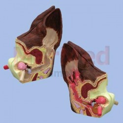
2-sided average size canine ear despicts...
2-sided average size canine ear despicts abnormal side illustrates inflamed inner ear structures, inflammatory exudates in tympanic bulla, ear canal with partial occlusion from cellular hyperplasia size 12 x 7 x 5 cm
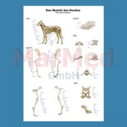
2 605,08 KčAnatomical wall chart: Canine skeleton...
Anatomical wall chart: Canine skeleton Laminated, 70 x 100 cm, with hanger and metal mounting rods. Languages: German, Latin, English
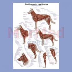
634,44 KčTeaching Chart: Canine Muscular Anatomy,...
Teaching Chart: Canine Muscular Anatomy, Laminated, 70 x 100 cm, with hanger and metal mounting rods. Languages: German, Latin, English
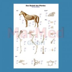
634,44 KčAnatomical wall chart: Horse skeleton...
Anatomical wall chart: Horse skeleton Laminated, 70 x 100 cm, with hanger and metal mounting rods. Languages: German, Latin, English
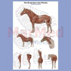
634,44 KčTeaching Chart: Equine Muscular Anatomy,...
Teaching Chart: Equine Muscular Anatomy, Laminated, 70 x 100 cm, with hanger and metal mounting rods. Languages: German, Latin, English
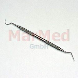
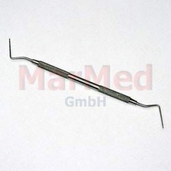
634,44 KčExplorer double ended one end rounded, one...
Explorer double ended one end rounded, one end double bent
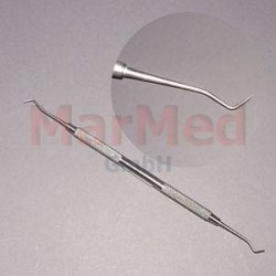
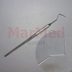
167,06 KčPeriodontometer Fox, single sided,...
Periodontometer Fox, single sided, 1-2-3-5-7-9-10-11 mm graduation
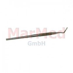
171,03 KčPeriodontometer, double ended, 1-10 and...
Periodontometer, double ended, 1-10 and 3,5-5,5-8,5-12,5 mm graduation
196,89 KčPeriodontometer WHO, single sided,...
Periodontometer WHO, single sided, 3,5-2-3-3 graduation
165,09 Kč


